Del Mar Photonics - Newsletter April 2011
Featured Application
Description of the application of multiphoton imaging from one of our potential customers
Diagnosing liver fibrosis: a new scoring method based on nonlinear microscopy
Liver fibrosis refers to the scarring response of the liver to external
aggressions, like hepatitis B or C viruses (HBV, HCV) infecting more than 500
million people worldwide or excessive alcohol intake. The over-accumulation of
fibrous tissue (mainly collagen) modifies liver function, and ultimately leads
to cirrhosis and life threatening complications such as primary liver cancer.
Cirrhosis is the leading cause for liver transplantation in France. In Rennes,
more than 3800 patients with HCV infection are treated in CHU Pontchaillou.
Thus, liver chronic diseases constitute an important public health issue, and it
is important to have a reliable diagnosis and early detection of fibrosis. Liver
biopsy is the gold standard for assessing liver fibrosis and cirrhosis but the
semi-quantitative scores used until now are inappropriate for accurate follow-up
(especially for the assessment of the effect of antifibrotic treatments) and are
subjected to possible intra and inter observer lecture variations. Therefore,
this study was motivated by the need to develop a robust scoring system for
diagnosing liver fibrosis/cirrhosis that avoids the risk of inter-observer
variations and allows for precise quantitative measurement of the amount of
collagen, as well as providing new informations on the structure of collagen
deposits and 3D reconstruction of the ECM network arrangement.
Nonlinear microscopy is a unique tool which provides intrinsic optical
sectioning and high in-depth imaging due to the inherent localization of the
nonlinear excitation at the objective focal volume, while drastically reducing
out-of-focus photobleaching and phototoxicity. Moreover, type-I or III collagen,
the main component of the fibrosis deposit in liver, can be selectively imaged
by the second harmonic generation (SHG) signals without staining. From this
specific contrast, we developed a new scoring method in assessing human liver
fibrosis [1]. This multidisciplinary work is an example of fruitful
collaboration between physicists (IPR/UMR CNRS 6251), biologists (EA
Seraic/IRSET, IFR140) and hepatologists and pathologists (CHU Pontchaillou).
Although SHG scoring may lead to a more accurate diagnosis of liver fibrosis,
our study also opens many perspectives [2]. In particular, we are working in
using the potentialities of 3D reconstruction of collagen networks, as well as
orientational patterns [3,4], to get a better insight into the collagen deposit
at various scales and fibrosis levels. Feasibility of endoscopic SHG exploration
for rodent models is also planned.
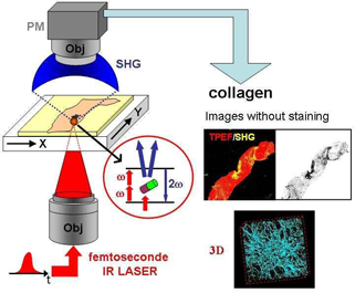
Legend: Nonlinear microscopy of human liver biopsies. Two-photon excitation
fluorescence (TPEF) and Second-harmonic generation (SHG) were acquired
simultaneously in 2D or 3D image stacks. SHG images reveal selectively fibrillar
collagen of type I or III without staining. A score proportional to the amount
of collagen deposit was derived from these images (Image courtesy of Universite
de Rennes 1).
Scientific contact
References:
[1] Gailhouste L., Le Grand Y., Odin C., Guyader D., Turlin B., Ezan F., Desille
Y., Guilbert T., Bessard A., Fremin C., Theret N., Baffet G., "Fibrillar
collagen scoring by second harmonic microscopy: A new tool in the assessement of
liver fibrosis." Journal of Hepatology, 52, (3), 398-406 (2010).
[2] Bedossa P., Editorial: "Harmony in liver fibrosis..." Journal of Hepatology,
52, (3), 313-314 (2010).
[3] Odin C., Guilbert T., Al Kilani A., Boryskina O.P., Fleury V., Le Grand Y.,
"Collagen and myosin characterization by orientation field second harmonic
microscopy.", Optics express, 16, (20), 16151-16165 (2008).
[4] Odin C., Le Grand Y., Renault A., Gailhouste L., Baffet G.,"Orientation
Fields of Non-Linear Biological Fibrils by Second Harmonic Generation
Microscopy." Journal of Microscopy, 229, 32-38 (2008).
Del Mar Photonics supplies multi-photon lasers and systems based on cost effective femtosecond sources:
Multiphoton Imaging
Related Del Mar Photonics products
If you can't find information about the products that you are looking for just e-mail our Sales Team and we'll e-mail or call you back right away!
Trestles LH femtosecond lasers with integrated DPSS DMPLH laser pump
Trestles LH10-fs/CW laser system at UC Santa Cruz Center of Nanoscale Optofluidics
Del Mar Photonics Tresltes LH laser used for STED microscopy of nanodiamons
Femtosecond Lasers
Trestles femtosecond Ti:Sapphire
laser
Trestles Finesse femtosecond
Ti:Sapphire laser with integrated DPSS
pump laser
Trestles LH femtosecond Ti:Sapphire laser with integrated DPSS pump laser
Trestles Opus femtosecond Ti:Sapphire laser with built in 3 Watt DPSS laser
Teahupoo Rider femtosecond
amplified Ti:Sapphire laser
Mavericks femtosecond
Cr:Forsterite laser
Tamarack femtosecond fiber laser
(Er-doped fiber)
Buccaneer femtosecond
OA fiber laser (Er-doped fiber) and SHG
Cannon Ultra-broadband
light source
Tourmaline femtosecond
Yt-doped fiber laser
Yb-based high-energy fiber laser system kit, model Tourmaline Yb-ULRepRate-07
Ytterbium-doped Femtosecond Solid-State Laser Tourmaline Yb-SS400